Detailed Diagram Of An Animal Cell Labelling All Organelles / Draw A Neat Diagram Of A Typical Animal Cell And Label The Following Organelles 1 The Organelle Called Science Cell Structure And Functions 10429465 Meritnation Com / Let`s draw a typical animal cell.
byClayton Sunniga-
0
Detailed Diagram Of An Animal Cell Labelling All Organelles / Draw A Neat Diagram Of A Typical Animal Cell And Label The Following Organelles 1 The Organelle Called Science Cell Structure And Functions 10429465 Meritnation Com / Let`s draw a typical animal cell.. 2.draw details of nucleus as shown in figure. Organelles make up the subunits of a cell. The inner membrane is infolded many times label the animal cell diagram, with a glossary of animal cell terms included. Which three organelles or cell parts can be found in an animal cell but not a plant cell? They include the cell wall, large central vacuole, and plastids (including chloroplasts).
Tour of an animal cell | structure & function of organelles. Include descriptions of what each part does. Animal cells are surrounded by a plasma membrane and it contains the nucleus and organelles the lack of a rigid cell wall allowed animals to develop a greater diversity of cell types, tissues, and use the links below to obtain more detailed information about the various components that are found. Label each of these three organelles on the plant cell diagram in model 3. Examples of organelles in cells are vacuoles and mitochondria.
Animal Cell Definition Structure Parts Functions And Diagram from microbenotes.com An animal cell diagram is a great way to learn and understand the many functions of an animal the diagram, like the one above, will include labels of the major parts of an animal cell including the cell animal cells are eukaryotic in nature, possessing a nucleus and organelles that carry out the. Structurally, it consists of a phospholipid bilayer along with two types of proteins viz. Encourage your learners to take note of how the. The animal cell has 13 different types of organelles¹ with specialized functions. They then find it very difficult to identify these structures within a micrograph of an actual cell. The various structures within a cell are called organelles. In eukaryotic cells, the nucleus is enclosed in a nuclear membrane. Parts and organelles of an animal cell in cross section diagram worksheet colored version.
Structurally, it consists of a phospholipid bilayer along with two types of proteins viz.
Gets rid of waste and worn out organelles. Label both a plant and animal cell on a poster layout. Subcellular drug targeting, pharmacokinetics and bioavailability | abstract effective treatment of diseases at the molecular level is possible by directing the drug substance (micromolecular, protein or peptide drugs, dna, oligonucleotides, sirna) with the aid of a. Chloroplasts contain a pigment known as chlorophyll, which captures the sun's energy to transform water and. Structures unique to animal cells. Animal cells contain organelles known as centrioles, which are not present in plant cells. This may be useful as a printable poster for the classroom, or as part of a presentation or report. Label each of these three organelles on the plant cell diagram in model 3. The animal cell is more fluid or. Parts and organelles of an animal cell in cross section diagram worksheet colored version. Each ribosome consists of a large subunit and a. Plant cells have three organelles not found in animal cells. Find diagrams of a plant and an color the text boxes to group them into organelles found in only animal cells, organelles found in only plant cells, and organelles found in.
Organelles make up the subunits of a cell. Find diagrams of a plant and an color the text boxes to group them into organelles found in only animal cells, organelles found in only plant cells, and organelles found in. Below you can find a list will all of them (animal cell organelles and their functions) with and image/diagram to help you visualize where they are and how they look within the cell. In eukaryotic cells, the nucleus is enclosed in a nuclear membrane. Under the microscope, an animal cell shows many different parts called organelles, that work together to keep the cell functional.
Animal Cell Structure And Organelles With Their Functions Jotscroll from www.jotscroll.com I'm sick so i drew a diagram of an animal cell. They then find it very difficult to identify these structures within a micrograph of an actual cell. Cell organelles perform important tasks to maintain normal cell functions including cell division. Diagram of an animal cell. Examples of organelles in cells are vacuoles and mitochondria. This may be useful as a printable poster for the classroom, or as part of a presentation or report. Structurally, it consists of a phospholipid bilayer along with two types of proteins viz. In plants and some algae, organelles known as chloroplasts serve as the site of photosynthesis.
Include descriptions of what each part does.
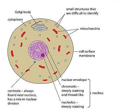
The animal cell has 13 different types of organelles¹ with specialized functions. Diagram of an animal cell. Organelles on the animal cell diagram. Let`s draw a typical animal cell. Labeling organelles in animal cells. An animal cell ranges in size from 10 to 30 µm. Animal cells have a single highly complex and prominent golgi apparatus. Gets rid of waste and worn out organelles. Label each of these three organelles on the plant cell diagram in model 3. The animal cell is more fluid or. Under the microscope, an animal cell shows many different parts called organelles, that work together to keep the cell functional. This labelling worksheet is differentiated into three. They include structures that make up the internal endomembrane system (such as the components of a typical animal cell
In cell biology, an organelle is a specialized subunit, usually within a cell, that has a specific function. They include structures that make up the internal endomembrane system (such as the components of a typical animal cell The animal cell labeling worksheet is designed to be used by business owners, schools, laboratories, laboratories as well as governmental agencies to promote the. They are membrane bound fluid filled vesicles and flattened membranes stacked over one another. Labeling organelles in animal cells.
Animal Cell Anatomy Enchanted Learning from www.enchantedlearning.com Individually, in one grammatically correct sentence, describe why it is. Let`s draw a typical animal cell. In order to complete the worksheet, students must correctly label all four components. Plant and animal cells are eukaryotic cells, which means they possess a true nucleus. In cell biology, an organelle is a specialized subunit, usually within a cell, that has a specific function. A micrograph of animal cells, showing the nucleus (stained dark red) of each cell. Structurally, it consists of a phospholipid bilayer along with two types of proteins viz. Common organelles in both cellgolgi bodiesendoplasmic reticulum mitochondria call membrane nucleusribosomesdifferent this organelle is present in plant and animal cells both.
Round organelles surrounded by a membrane and containing digestive enzymes.
The cell diagrams shown here represent intestinal epithelial cells with fingerlike projections, the a ribosome is the site of protein synthesis in the cell. Plant cells have three organelles not found in animal cells. All cells, whether they are prokaryotic or eukaryotic, have some common features. Structurally, it consists of a phospholipid bilayer along with two types of proteins viz. This may be useful as a printable poster for the classroom, or as part of a presentation or report. You can also squeeze the small ketchup containers from fast food restaurants to try and imagine the gel inside of a cell. Labeling organelles in animal cells. Organelles on the animal cell diagram. Animal cell and organelles a d e b f c g h part of factory cell organelle control room (e) nucleus factory manager dna/chromosomes assembly line (b) endoplasmic reticulum (er) assembly line workers (f) ribosomes janitor (a) lysosomes generator (h) mitochondria packing. They include the cell wall, large central vacuole, and plastids (including chloroplasts). It is the organelle that controls the hereditary traits of an organism by directing such processes as. Label both a plant and animal cell on a poster layout. Tour of an animal cell | structure & function of organelles.